Delivery of Cas9/sgRNA mixed with 306-O12B into adult mouse cochlea via cochleostomy
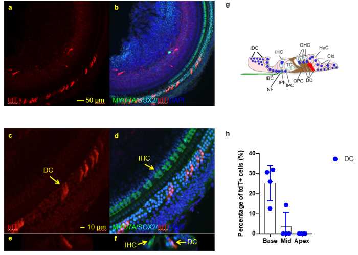
Delivery of Cas9/sgRNA mixed with 306-O12B into adult mouse cochlea via cochleostomy. a-f. Whole mount immunofluorescence staining of the base region of the cochlea using MYO7A (green) and SOX2 (cyan). Cochlea tissues were collected 3-7 days post injection. tdT signals were observed mainly in Deiters cells. (a-b), low magnification images; (c-d), high magnification images. (e-f) cross section view of (c-d). G, illustration of cells types with tdT signal in cochlea. H, quantification of tdT+ Deiters cells in cochlea. N=4, mean +/- SEM. DC, Deiters cells; IHC, inner hair cells