| Testing new LNPs (lipid nanoparticles) for delivery of Cas9 mRNA/sgRNA in adult mouse cochlea |
| 113-O12B |
| Editing efficiency is calculated by percentage of tdTomato positive cells |
| base of cochlear canal |
| cochlear inner hair cell |
| Editing Efficiency |
| 15000000597 |
|
| CleanCap® Cas9 |
| 113-O12B |
| Nanoparticle |
| sg298 |
|
|
| Mouse |
| B6.Cg-Gt(ROSA)26Sor^tm14(CAG-tdTomato)Hze/J |
|
| 4-8 weeks |
|
| cochleostomy |
| cochlea |
| LNP/Cas9/sgRNA=0.15/0.01/0.01ug |
| once |
| 3 nl/sec |
| 300 nanoliters |
| 5 days |
| mRNA |
|
|
|
| AB_10015251 |
| 25-6790 |
| Anti-Myosin VIIa antibody, Proteus Biosciences |
|
| AB_2286684 |
| sc-17320 |
| Sox-2 (Y-17) Antibody, Santa Cruz Biotechnology |
|
| Measured Values |
| Samples Size: | 2 |
| |
| Mean |
4.76 |
| 1 |
9.52 |
| 2 |
0 |
| |
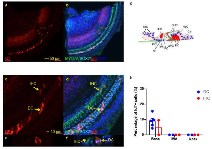
| Delivery of Cas9/sgRNA mixed with 113-O12B into adult mouse cochlea via cochleostomy |
|
|
Delivery of Cas9/sgRNA mixed with 113-O12B into adult mouse cochlea via cochleostomy. a-f. Whole mount immunofluorescence staining of the base region of the cochlea using MYO7A (green) and SOX2 (cyan). Cochlea tissues were collected 5 days post injection. tdT signals were observed mainly in Deiters cells. (a-b), low magnification images; (c-d), high magnification images. (e-f) cross section view of (c-d). G, illustration of cells types with tdT signal in cochlea. H, quantification of tdT+ Deiters cells in cochlea. N=2, mean +/- SEM. DC, Deiters cells; IHC, inner hair cells. |
|