Development System Testing Shuttle Peptides ability to deliver GFP-NLS to airway epithelia. | SCGE Toolkit Delivery of GFP via shuttle peptides to mouse airway epithelium via nasal instilation. Delivery efficiency was quantified in large and small airways by counting the number of GFP positive cells divided by the number of DAPI cells.
Experiment: Testing Shuttle Peptides ability to deliver GFP-NLS to airway epithelia.
PI:
Paul B McCray, Jr, MD
Description: Delivery of GFP via shuttle peptides to mouse airway epithelium via nasal instilation. Delivery efficiency was quantified in large and small airways by counting the number of GFP positive cells divided by the number of DAPI cells.
Delivery Assays:
Quantification of GFP+ cells in large and small airways following 1 delivery of GFP protein by GFP positive cells compared to DAPI stained cells.
Select experimental variable to highlight records on the chart:
Note: Hover over the bars to view additional information
Loading...
Results
SCGE Toolkit downloaded on: 2026/07/10 02:16:26; Please cite the Somatic Cell Genome Editing Consortium Toolkit NIH HG010423 when using publicly accessible data in formal presentation or publication. SCGE Experment ID: 18000000002. PI:
Paul B McCray Jr MD
Engineered amphiphilic peptides enable delivery of proteins and CRISPR-associated nucleases to airway epithelia. NCBI
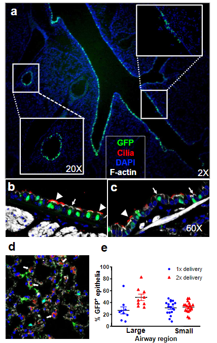
GFP-NLS protein delivery to mouse airways using S10 peptide. a Fluorescence image of lung tissue section 18 h following two intranasal doses of [S10]: 40μM; [GFP]: 20μMin50μl; ×2 magnification. Insets show the large and small airways at ×20 magnification. b,c Localization of GFP in different cell types. GFP co-localized with specific markers of cilia (α-tubulin, red), F-actin (phalloidin stain, gray), and nuclei (DAPI, blue) in large (b) and small airway epithelia (c). Non-ciliated cells were identified by the absence of α-tubulin staining; ×40 magnification. Arrowheads indicate ciliated cells (α-tubulin); arrows indicate non-ciliated cells. n=4 mice per group. d GFP localization in distal lung region. Co-localization of GFP and SP-C (red), a marker of alveolar type II cells, F-actin (phalloidin stain, gray), and nuclei (DAPI, blue); ×40 magnification. White arrows indicate co-localization of GFP and SP-C. e Quantitation of GFP+ cells in large and small airways following 1 or 2 deliveries of GFP protein. Results are presented as mean ± SE; n=4 mice per group. Pubmed Link