Experiment: Testing virus region 8 (VR8) mutant cross-species compatible Adeno Associated Viruses (ccAAVs) in mice.
PI: Aravind Asokan, PhD
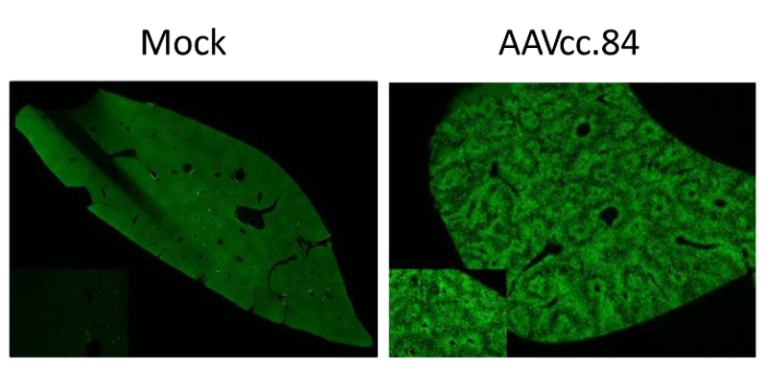
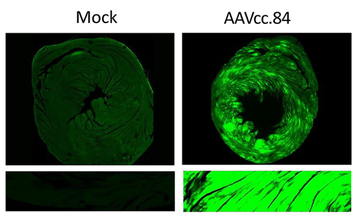
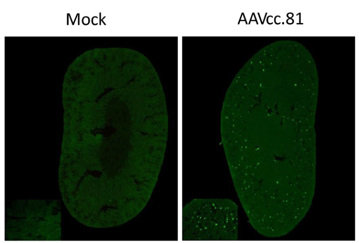
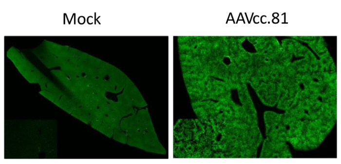
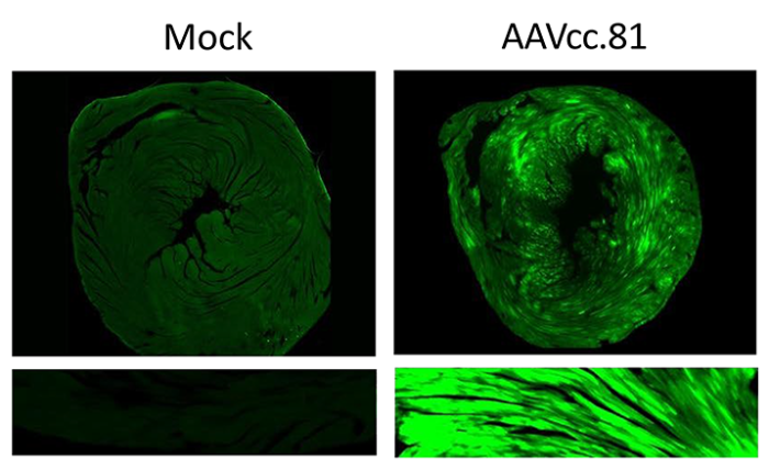
- Native fluorescence was quantified using Image J. For each animal, 6 images were taken from 2 sections and used to calculate corrected total cell fluorescence using the formula: Integrated Density – (Area of selected image X Mean fluorescence of background readings).
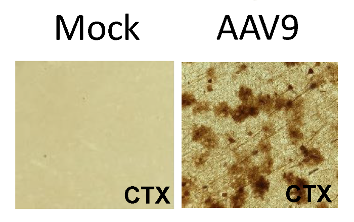
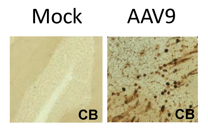
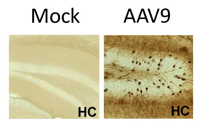
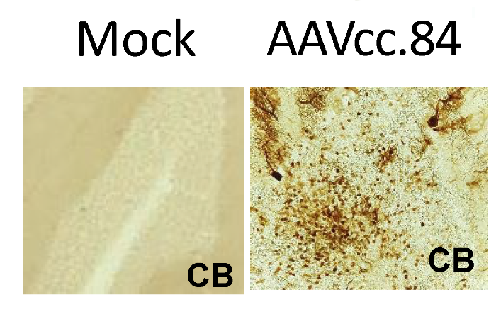
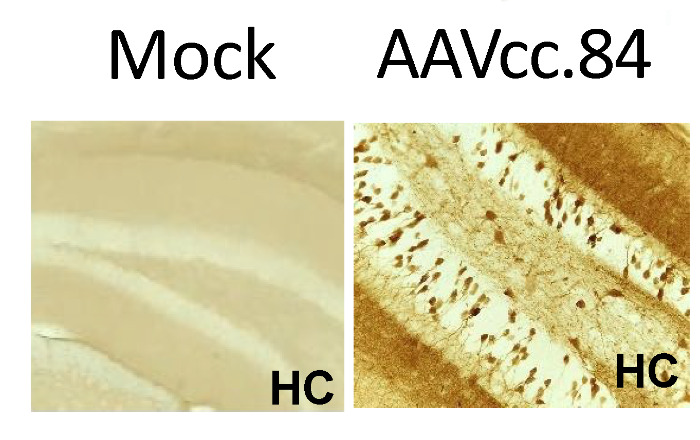
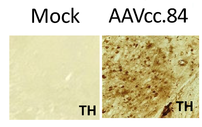
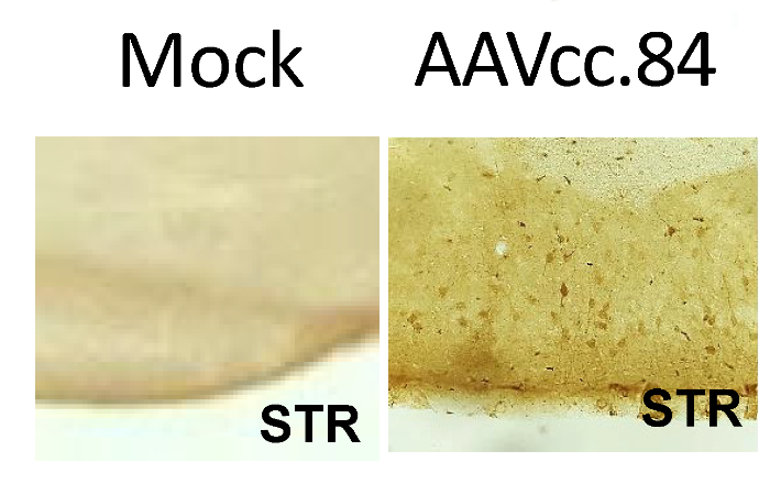
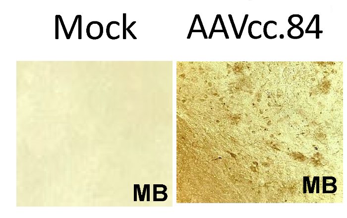
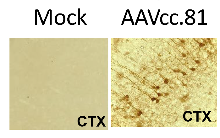
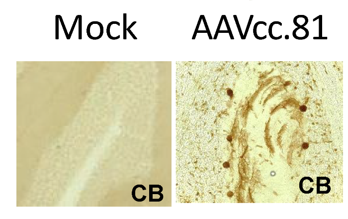
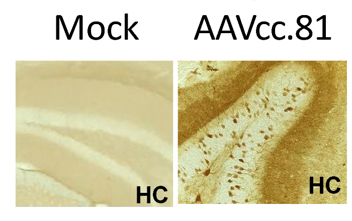
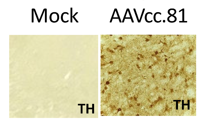
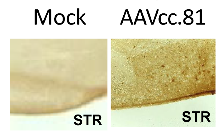
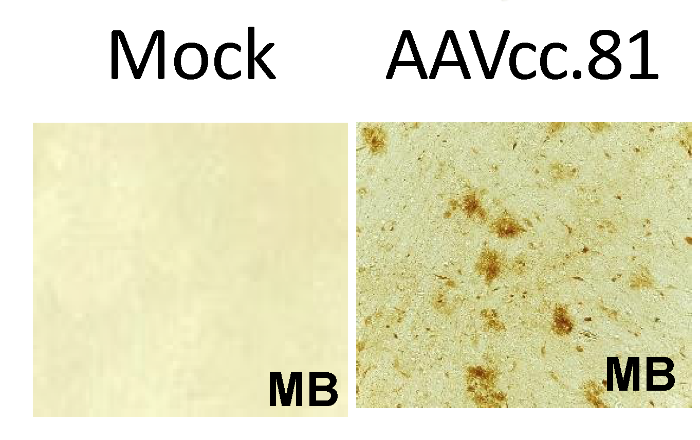
- Relative neuronal transduction levels were quantified by counting the number of transduced neurons, identified based on morphology, for each brain region per 50um sagittal section
-
Other experiments in this project: 6
- Testing virus region 4 (VR4) mutant cross-species compatible Adeno Associated Viruses (ccAAVs) in mice.
- Cre Recombinase dose escalation study in Ai9 mice
- Comparing CRISPR/Cas9 gene editing efficencies between AAV9 and AAVcc47 in Ai9 mice with a 1:1 Cas9 to sgRNA ratio (CB promoter)
- Comparing CRISPR/Cas9 gene editing efficencies between AAV9 and AAVcc47 in Ai9 mice with a 1:1 cas9 to sgRNA ratio (CMV promoter)
- Comparing CRISPR/Cas9 gene editing efficencies between AAV9 and AAVcc47 in Ai9 mice with a 1:1 Cas9 to sgRNA ratio (CMV promoter) and self complementary sgRNA vector.
- Comparing CRISPR/Cas9 gene editing efficiencies between AAV9 and AAVcc47 in Ai9 mice with a 1:3 Cas9 to sgRNA ratio (CMV promoter)
Note: Hover over the bars to view additional information
Results |
| Delivery Efficiency | |||||||||||
|---|---|---|---|---|---|---|---|---|---|---|---|
| Condition | Tissue | Cell Type | Sex | Age | Model | Vector | Dosage | Injection Frequency | # of cells in 50um Section | Corrected Total Fluorescence | Image |
| AAV9 GFP | cerebral cortex | neuron | Male | 8 weeks | C57BL/6 mouse (Asokan study) | AAV9-GFP | 5e13 vg/kg | once |
 |
||
| AAV9 GFP | cerebral cortex | astrocyte | Male | 8 weeks | C57BL/6 mouse (Asokan study) | AAV9-GFP | 5e13 vg/kg | once |
 |
||
| AAV9 GFP | cerebellum | neuron | Male | 8 weeks | C57BL/6 mouse (Asokan study) | AAV9-GFP | 5e13 vg/kg | once |
 |
||
| AAV9 GFP | cerebellum | astrocyte | Male | 8 weeks | C57BL/6 mouse (Asokan study) | AAV9-GFP | 5e13 vg/kg | once |
 |
||
| AAV9 GFP | hippocampal formation | neuron | Male | 8 weeks | C57BL/6 mouse (Asokan study) | AAV9-GFP | 5e13 vg/kg | once |
 |
||
| AAV9 GFP | hippocampal formation | astrocyte | Male | 8 weeks | C57BL/6 mouse (Asokan study) | AAV9-GFP | 5e13 vg/kg | once |
 |
||
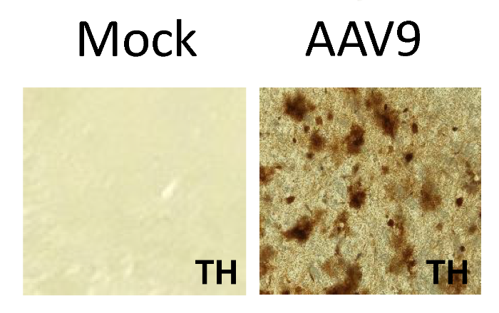
| AAV9 GFP | dorsal plus ventral thalamus | neuron | Male | 8 weeks | C57BL/6 mouse (Asokan study) | AAV9-GFP | 5e13 vg/kg | once |
 |
||
| AAV9 GFP | dorsal plus ventral thalamus | astrocyte | Male | 8 weeks | C57BL/6 mouse (Asokan study) | AAV9-GFP | 5e13 vg/kg | once |
 |
||
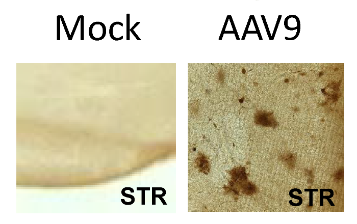
| AAV9 GFP | striatum | neuron | Male | 8 weeks | C57BL/6 mouse (Asokan study) | AAV9-GFP | 5e13 vg/kg | once |
 |
||
| AAV9 GFP | striatum | astrocyte | Male | 8 weeks | C57BL/6 mouse (Asokan study) | AAV9-GFP | 5e13 vg/kg | once |
 |
||
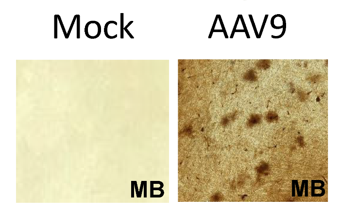
| AAV9 GFP | midbrain | neuron | Male | 8 weeks | C57BL/6 mouse (Asokan study) | AAV9-GFP | 5e13 vg/kg | once |
 |
||
| AAV9 GFP | midbrain | astrocyte | Male | 8 weeks | C57BL/6 mouse (Asokan study) | AAV9-GFP | 5e13 vg/kg | once |
 |
||
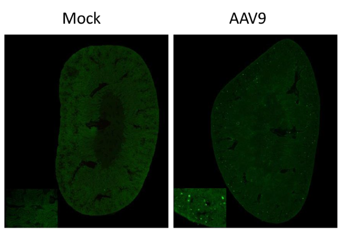
| AAV9 GFP | kidney | Male | 8 weeks | C57BL/6 mouse (Asokan study) | AAV9-GFP | 5e13 vg/kg | once |
 |
|||
| AAV9 GFP | liver | Male | 8 weeks | C57BL/6 mouse (Asokan study) | AAV9-GFP | 5e13 vg/kg | once |
 |
|||
| AAV9 GFP | heart | Male | 8 weeks | C57BL/6 mouse (Asokan study) | AAV9-GFP | 5e13 vg/kg | once |
 |
|||
| AAV9 GFP | skeletal muscle tissue | Male | 8 weeks | C57BL/6 mouse (Asokan study) | AAV9-GFP | 5e13 vg/kg | once |
 |
|||
| AAVcc84 | cerebral cortex | neuron | Male | 8 weeks | C57BL/6 mouse (Asokan study) | AAVcc84-GFP | 5e13 vg/kg | once |
 |
||
| AAVcc84 | cerebral cortex | astrocyte | Male | 8 weeks | C57BL/6 mouse (Asokan study) | AAVcc84-GFP | 5e13 vg/kg | once |
 |
||
| AAVcc84 | cerebellum | neuron | Male | 8 weeks | C57BL/6 mouse (Asokan study) | AAVcc84-GFP | 5e13 vg/kg | once |
 |
||
| AAVcc84 | cerebellum | astrocyte | Male | 8 weeks | C57BL/6 mouse (Asokan study) | AAVcc84-GFP | 5e13 vg/kg | once |
 |
||
| AAVcc84 | hippocampal formation | neuron | Male | 8 weeks | C57BL/6 mouse (Asokan study) | AAVcc84-GFP | 5e13 vg/kg | once |
 |
||
| AAVcc84 | hippocampal formation | astrocyte | Male | 8 weeks | C57BL/6 mouse (Asokan study) | AAVcc84-GFP | 5e13 vg/kg | once |
 |
||
| AAVcc84 | dorsal plus ventral thalamus | neuron | Male | 8 weeks | C57BL/6 mouse (Asokan study) | AAVcc84-GFP | 5e13 vg/kg | once |
 |
||
| AAVcc84 | dorsal plus ventral thalamus | astrocyte | Male | 8 weeks | C57BL/6 mouse (Asokan study) | AAVcc84-GFP | 5e13 vg/kg | once |
 |
||
| AAVcc84 | striatum | neuron | Male | 8 weeks | C57BL/6 mouse (Asokan study) | AAVcc84-GFP | 5e13 vg/kg | once |
 |
||
| AAVcc84 | striatum | astrocyte | Male | 8 weeks | C57BL/6 mouse (Asokan study) | AAVcc84-GFP | 5e13 vg/kg | once |
 |
||
| AAVcc84 | midbrain | neuron | Male | 8 weeks | C57BL/6 mouse (Asokan study) | AAVcc84-GFP | 5e13 vg/kg | once |
 |
||
| AAVcc84 | midbrain | astrocyte | Male | 8 weeks | C57BL/6 mouse (Asokan study) | AAVcc84-GFP | 5e13 vg/kg | once |
 |
||
| AAVcc84 | liver | Male | 8 weeks | C57BL/6 mouse (Asokan study) | AAVcc84-GFP | 5e13 vg/kg | once |
 |
|||
| AAVcc84 | heart | Male | 8 weeks | C57BL/6 mouse (Asokan study) | AAVcc84-GFP | 5e13 vg/kg | once |
 |
|||
| AAVcc81 | cerebral cortex | neuron | Male | 8 weeks | C57BL/6 mouse (Asokan study) | AAVcc81-GFP | 5e13 vg/kg | once |
 |
||
| AAVcc81 | cerebral cortex | astrocyte | Male | 8 weeks | C57BL/6 mouse (Asokan study) | AAVcc81-GFP | 5e13 vg/kg | once |
 |
||
| AAVcc81 | cerebellum | neuron | Male | 8 weeks | C57BL/6 mouse (Asokan study) | AAVcc81-GFP | 5e13 vg/kg | once |
 |
||
| AAVcc81 | cerebellum | astrocyte | Male | 8 weeks | C57BL/6 mouse (Asokan study) | AAVcc81-GFP | 5e13 vg/kg | once |
 |
||
| AAVcc81 | hippocampal formation | neuron | Male | 8 weeks | C57BL/6 mouse (Asokan study) | AAVcc81-GFP | 5e13 vg/kg | once |
 |
||
| AAVcc81 | hippocampal formation | astrocyte | Male | 8 weeks | C57BL/6 mouse (Asokan study) | AAVcc81-GFP | 5e13 vg/kg | once |
 |
||
| AAVcc81 | dorsal plus ventral thalamus | neuron | Male | 8 weeks | C57BL/6 mouse (Asokan study) | AAVcc81-GFP | 5e13 vg/kg | once |
 |
||
| AAVcc81 | dorsal plus ventral thalamus | astrocyte | Male | 8 weeks | C57BL/6 mouse (Asokan study) | AAVcc81-GFP | 5e13 vg/kg | once |
 |
||
| AAVcc81 | striatum | neuron | Male | 8 weeks | C57BL/6 mouse (Asokan study) | AAVcc81-GFP | 5e13 vg/kg | once |
 |
||
| AAVcc81 | striatum | astrocyte | Male | 8 weeks | C57BL/6 mouse (Asokan study) | AAVcc81-GFP | 5e13 vg/kg | once |
 |
||
| AAVcc81 | midbrain | neuron | Male | 8 weeks | C57BL/6 mouse (Asokan study) | AAVcc81-GFP | 5e13 vg/kg | once |
 |
||
| AAVcc81 | midbrain | astrocyte | Male | 8 weeks | C57BL/6 mouse (Asokan study) | AAVcc81-GFP | 5e13 vg/kg | once |
 |
||
| AAVcc81 | kidney | Male | 8 weeks | C57BL/6 mouse (Asokan study) | AAVcc81-GFP | 5e13 vg/kg | once |
 |
|||
| AAVcc81 | liver | Male | 8 weeks | C57BL/6 mouse (Asokan study) | AAVcc81-GFP | 5e13 vg/kg | once |
 |
|||
| AAVcc81 | heart | Male | 8 weeks | C57BL/6 mouse (Asokan study) | AAVcc81-GFP | 5e13 vg/kg | once |
 |
|||
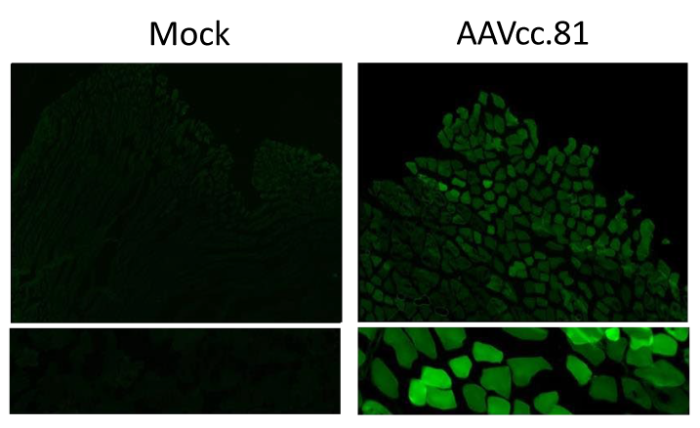
| AAVcc81 | skeletal muscle tissue | Male | 8 weeks | C57BL/6 mouse (Asokan study) | AAVcc81-GFP | 5e13 vg/kg | once |
 |
|||
| Expression of VR8 ccAAVs in the brain following IV injection |
|
|
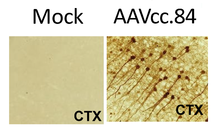
Expression of VR8 ccAAVs in the brain. Adult C57/B6 mice were injected intravenously at 5e13 vg/kg (N=3). A self-complementary GFP cassette was packaged into lead ccAAV mutants driven by a chicken-beta actin human hybrid (CBh) promoter. Mice were sacrificed 4-weeks post injection brain was post-fixed in 4% PFA for 24 hrs. Tissue was cut into 50mm sagittal sections using a vibratome. Immunohistochemical staining of brain sections revealed neuronal and astrocytic expression (representative images are shown) |
| Expression of VR8 ccAAVs in cardiac and skeletal muscle |
|
|
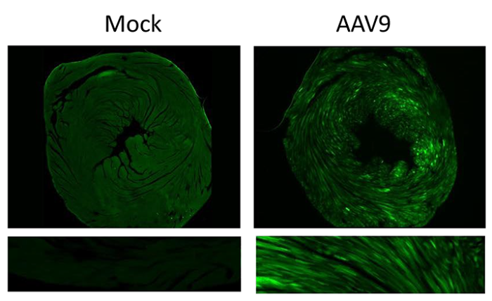
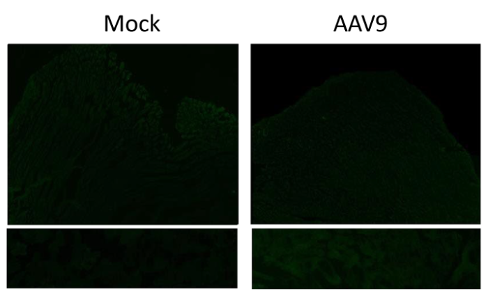
Expression of VR8 ccAAVs in cardiac and skeletal muscle. Adult C57/B6 mice were injected intravenously at 5e13 vg/kg (N=3). A self-complementary GFP cassette was packaged into lead ccAAV mutants driven by a chicken-beta actin human hybrid (CBh) promoter. Mice were sacrificed 4-weeks post injection and heart and skeletal muscle was post-fixed in 4% PFA for 24 hrs. Fixed tissue was embedded in agarose and cut into 50mm thick sections using a vibratome. Native fluorescence was quantified using Image J. For each animal, 6 images were taken from 2 sections and used to calculate corrected total cell fluorescence using the formula: Integrated Density (Area of selected image X Mean fluorescence of background readings). |
| Expression of VR8 ccAAVs in liver and kidney |
|
|
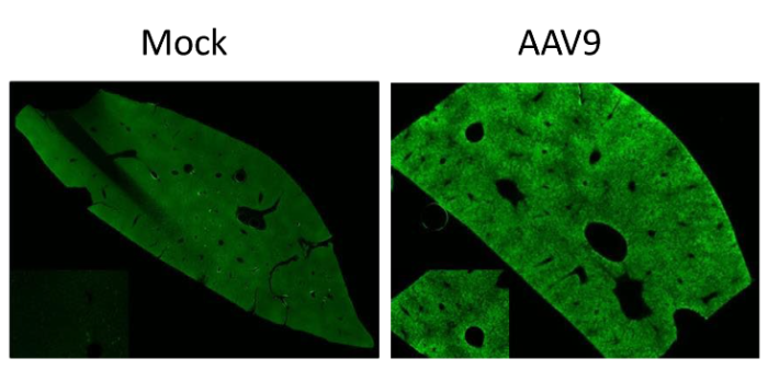
Expression of VR8 ccAAVs in liver and kidney. Adult C57/B6 mice were injected intravenously at 5e13 vg/kg (N=3). A self-complementary GFP cassette was packaged into lead ccAAV mutants driven by a chicken-beta actin human hybrid (CBh) promoter. Mice were sacrificed 4-weeks post injection and liver and kidney was post-fixed in 4% PFA for 24 hrs. Fixed tissue was embedded in agarose and cut into 50mm thick sections using a vibratome. Native fluorescence was quantified using Image J. For each animal, 6 images were taken from 2 sections and used to calculate corrected total cell fluorescence using the formula: Integrated Density (Area of selected image X Mean fluorescence of background readings). |
