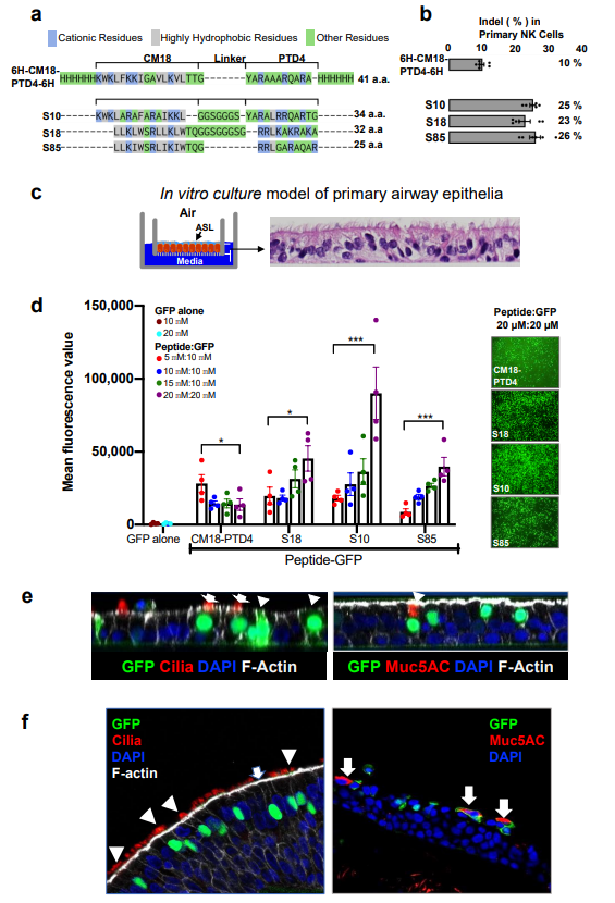
Shuttle peptide design and protein delivery to airway epithelia. A) Amino acid sequences of shuttle peptides. Sequences aligned to highlight structural similarities. Cationic residues are highlighted in blue; hydrophobic residues are in gray. Remaining residues are in green. B) Indel% in primary NK cells following Cas12a RNP delivery targeting NKG2A gene with indicated shuttle peptide ([Cas12a]: 1.33 uM; [crRNA]: 2.0 µM). Results quantified 48 h after delivery (mean ± SE; n = 3 independent experiments). C) Shuttle peptide-mediated delivery of nuclear-targeted GFP protein to human airway epithelia (HAE) and tracheal explants. Left panel: Schematic of primary HAE grown at airâliquid interface. ASL: airway surface liquid. Right panel: photomicrograph of well-differentiated primary HAE culture demonstrating pseudostratified columnar epithelium. Hematoxylin and eosin stain, Ã40 magnification. D) Efficiency of GFP delivery by different shuttle peptides. Peptides applied at four concentrations (5, 10, 15, or 20 uM); GFP concentrations 10 or 20 uM. Left graph: Cells were transduced with indicated peptide and GFP combinations and GFP quantified by fluorescence reader. Results mean ± SE; n = 4 donors. *P < 0.05, **P < 0.005, ***P < 0.001, by one-way ANOVA with Tukeyâs multiple comparison test. Right panels: Representative en face views of epithelial sheets following GFP delivery at the highest peptide:GFP concentration (20 uM:20 uM), Ã20 magnification. Formulations were applied for 15 min. E) GFP localization in multiple epithelial cell types following shuttle peptide-mediated delivery. HAEs were transduced for 15 min with GFP protein and S10 peptide. Left panel: confocal image (XZ) shows co-localization of GFP and specific marker of ciliated cells (α-tubulin, red, indicated by white arrows), nonciliated cells (white arrowheads), F-actin (phalloidin stain, gray), nuclei (DAPI, blue). Right panel: co-localization of GFP and goblet cell marker (Muc5AC, red, indicated by white arrowhead), nuclei (DAPI, blue), and F-actin (phalloidin stain, gray). n ⥠6 different donor epithelia; Ã64 magnification. F) Shuttle peptide delivery of GFP to human tracheal explants. S10 (20 uM) and GFP protein (20 uM) applied to tracheal explant for 1 h. Left panel: both ciliated (αtubulin, red, white arrowhead) and non-ciliated (white arrow) cells were transduced. Right panel: non-ciliated goblet cells were transduced (Muc5AC, red, white arrows); Ã40 magnification. https://doi.org/10.1038/s41467-019-12922-y