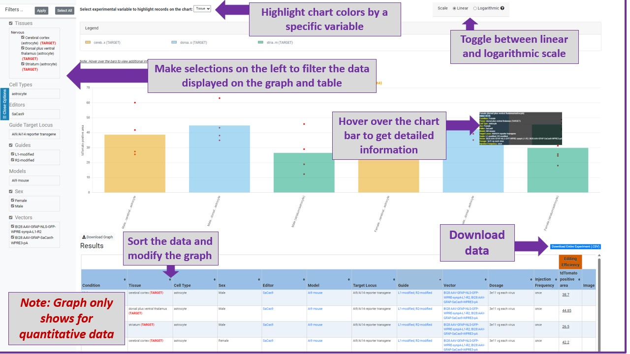
Note: Hover over the bars to view additional information
SCGE Toolkit downloaded on: 2026/06/13 09:27:18; Please cite the Somatic Cell Genome Editing Consortium Toolkit NIH HG010423 when using publicly accessible data in formal presentation or publication. SCGE Experment ID: 18000000010. PI:
Paul B McCray Jr MD
| Publication Title |
|
Engineered amphiphilic peptides enable delivery of proteins and CRISPR-associated nucleases to airway epithelia. NCBI |
| S10 peptide delivery of Cas9 RNP shows editing in ROSAmT/mG locus in vivo |
|
|
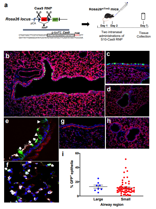
S10 peptide delivery of Cas9 RNP shows editing in ROSAmT/mG locus in vivo. Cas9 RNP directed to loxP sites flanking the tdTomato cassette were administered with S10 peptide once daily on 2 consecutive days. Seven days later, conversion of tdTomato to GFP expression was visualized in lung tissue sections. A) Schematic of gRNA targeting two loxP sequences flanking tdTomato gene and experimental protocol for Cas9 RNP delivery to ROSAmT/mG mice. B) Fluorescence image of large airway 7 days following two intranasal doses of [S10]: 40 µM; [Cas9]: 1.33 µM; [gRNA]: 2 µM; Ã2 magnification. GFP expression denotes edited cells. C) Editing in a large airway, Ã20 magnification. D) Editing in a small airway; Ã20 magnification. E) Co-localization of GFP and marker of ciliated cells (α-tubulin, white) in large airway. Arrows indicate α-tubulin co-localization with GFP. Arrowheads denote edited (GFP+) nonciliated cells negative for α-tubulin; Ã40 magnification. F) Co-localization of GFP+ and SP-C (white) identifies alveolar type II cells (arrows) in distal lung; Ã40 magnification. G) Representative image of large airway after delivery of Cas9 RNP alone shows no editing, Ã20 magnification. H) Representative image of small airways after delivery of Cas9 RNP alone shows no editing; Ã20 magnification. I) Editing efficiency of Cas9 in large and small airways quantified by the number of GFP+ cells. Horizontal lines indicate mean ± SE; n = 5 mice/group. Pubmed |
| S10 peptide delivery of Cas12a RNP shows editing in ROSAmT/mG locus in vivo |
|
|
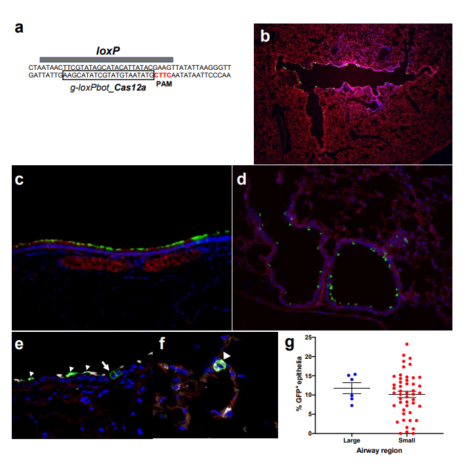
S10 peptide delivery of Cas12a RNP shows editing in ROSAmT/mG locus in vivo. A) Schematic of Cas12a gRNA targeting both loxP sequences flanking tdTomato gene in ROSAmT/mG mice. S10 peptide and Cas12a RNP (2.5 uM Cas12a; 2.0 uM gRNA) directed to suboptimal PAM target in loxP sites flanking the tdTomato cassette administered twice a day for 2 days, via intranasal instillation. Seven days later, conversion of tdTomato to GFP expression was visualized in lung tissue sections. B) Cas12a mediated deletion of tdTomato in ROSAmT/mG in large airway epithelia in vivo; x2 magnification. C) Editing in a large airway; x20 magnification. D) Editing in a small airway; x20 magnification. E) Co-localization of GFP and marker of ciliated cells (a-tubulin, white) in large airway. Arrowheads indicate GFP and a-tubulin co-localization. Arrow denotes edited (GFP+) non-ciliated cell negative for a±-tubulin; x40 magnification. F) Distal lung region. Co-localization of GFP+ and SP-C (white) identifies alveolar type II cells (arrowhead); x40 magnification. G) Editing efficiency of Cas12a RNP in large and small airways quantified by the number of GFP+ cells. Horizontal lines indicate mean ± SE; n = 3 mice/group. https://doi.org/10.1038/s41467-019-12922-y |
× Close Overview